A Levels Biology (9700)•9700/12/O/N/22
Explanation
Significant figures in microscope field of view area calculation
Steps:
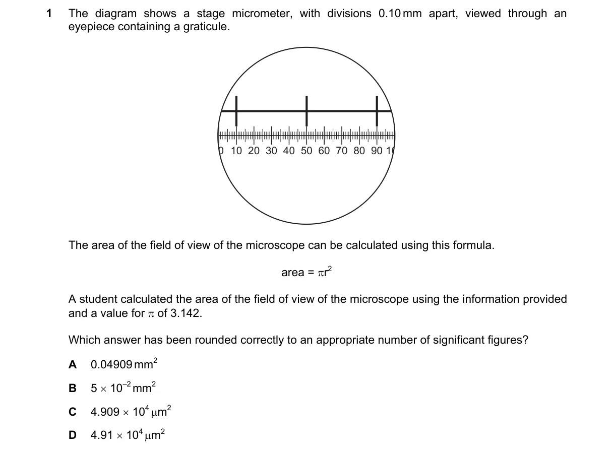
- Calibrate eyepiece graticule against stage micrometer: each 0.1 mm division corresponds to a measured number of graticule units, giving scale factor with 3 significant figures (e.g., diameter ≈ 1.25 mm or 1250 μm).
- Determine radius r from half the calibrated diameter, preserving 3 significant figures.
- Compute area = π r² with π = 3.142 (4 significant figures); result has 3 significant figures due to limiting measurement precision.
- Convert to μm² if needed (1 mm = 1000 μm, exact) and round to 3 significant figures: 491 μm².
Why D is correct:
- 491 has 3 significant figures, matching the precision of 0.1 mm divisions (2–3 figures) limited by calibration accuracy.
Why the others are wrong:
- A: 2 significant figures and incorrect magnitude/units (too small for typical field).
- B: Only 2 significant figures; incomplete (missing m value).
- C: 3 significant figures but incorrect scientific notation (missing exponent) and wrong magnitude.
Final answer: D
Topic: The microscope in cell studies
Practice more A Levels Biology (9700) questions on mMCQ.me